BOOK AN APPOINTMENT-> click here
LIST OF INSURANCE ACCEPTED-> click here
DAILY ACTIVITIES-> click here
ANY COMMENTS-> click here
MASTER HEALTH PACKAGES-> click here
PATIENT FEEDBACK

2D ECHO

logo

Procedures and treatment

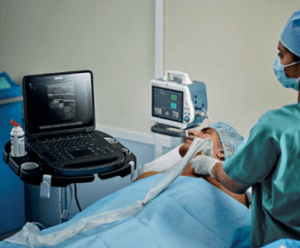
  • A standard echo doesn't require any special preparations or followup. If you're having a TEE, you usually shouldn't eat or drink for 8 hours prior to the test. During a standard echo, your doctor or sonographer will move a wand-like device called a transducer around on your chest to get pictures of your heart.
  • Normal Ranges for LV Size and Function Normal values for LV chamber dimensions (linear), volumes and ejection fraction vary by gender. A normal ejection fraction is 53-73% (52-72% for men, 54-74% for women).

Echocardiography (echo) shows the size, structure, and movement of various parts of your heart. These parts include the heart valves, the septum (the wall separating the right and left heart chambers), and the walls of the heart chambers. Doppler ultrasound shows the movement of blood through your heart.

2D Echocardiography or 2D Echo of heart is a test in which ultrasound technique is used to take pictures of heart. It displays a cross sectional 'slice' of the beating heart, showing chambers, valves and the major blood vessels of heart.